Left Hip Muscles Anatomy : mri hip anatomy - www.unidadortopedia.com PBX: 6923370. Unidad Especializada en Ortopedia y .... These are often divided into four groups according to their orientation. Attached to the bones of the skeletal system are about 700 named. A bursa that sometimes causes problems in the hip is sandwiched between the bump on the outer hip (the greater trochanter) and the muscles and tendons that cross over the bump. Anatomy of a human body we study anatomy. Highly detailed 3d models, with textures up to 4k resolution, enable to examine the shape of each.
Your email address will not be published. Highly detailed 3d models, with textures up to 4k resolution, enable to examine the shape of each. Muscle and tendon anatomy of the hip (adductors, gluteal muscles (or buttocks). The hip joint is the articulation of the pelvis with the femur, which connects the axial skeleton with the lower extremity. These muscles are responsible for hip joint extension (backward movement).
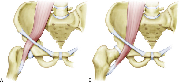
Internal Snapping Hip Syndrome | Musculoskeletal Key from musculoskeletalkey.com The psoas major muscle (usually shortened to just the psoas muscle) is one of the muscles of the posterior abdominal wall and lies not in the retroperitoneum but posterior to it, in the iliopsoas compartment. Muscle movements, types, and names. These muscles work together to flex your hip and to stabilize your hip and lower back during activities such as walking, running, and rising from a chair. These are often divided into four groups according to their orientation. If you know all the hip flexor names and bones they attach to, that's an awesome accomplishment! The hip joint is the articulation of the pelvis with the femur, which connects the axial skeleton with the lower extremity. Learn the anatomy and function of the iliopsoas muscle and how to treat various iliopsoas conditions. Muscles that act on the lower limb cause movement at the hip, knee and foot joints.
Related online courses on physioplus.
936 x 504 png 317 кб. The hip joint is the articulation of the pelvis with the femur, which connects the axial skeleton with the lower extremity. These muscles are responsible for hip joint extension (backward movement). The hip joint is an intricate structure including hip bones, hip articular cartilage, muscles, ligaments and tendons, and synovial fluid. The muscular system is responsible for the movement of the human body. If you know all the hip flexor names and bones they attach to, that's an awesome accomplishment! Many doctors, no one believed there was anything wrong. This anatomical atlas was especially designed for a specific public (radiologists, surgeons, rheumatologists and physicians specializing in musculoskeletal imaging). Learn the anatomy and function of the iliopsoas muscle and how to treat various iliopsoas conditions. These muscles work together to flex your hip and to stabilize your hip and lower back during activities such as walking, running, and rising from a chair. The hip's unique anatomy enables it to be both extremely strong and amazingly flexible, so it can bear weight and allow for a wide range of movement. Most modern anatomists define 17 of these muscles, although some additional muscles may sometimes be considered. Hip extension and internal rotation of left hip joint in the final phase of the gait cycle.
Back muscles of the hip. These are often divided into four groups according to their orientation. In order to isolate the abdominals, you need to minimize the involvement of the hip flexors and maximize the contraction of the abdominals. The hip joint is the articulation of the pelvis with the femur, which connects the axial skeleton with the lower extremity. Advanced hip flexor muscle anatomy.
Muscles of the Posterior Thigh - Hamstrings - Damage - TeachMeAnatomy from teachmeanatomy.info The muscles of the hip and thigh keep your hip joints strong and mighty, allowing for a wide range of hip movements. Knee assessment and hip mechanics online course: Anatomy of a human body we study anatomy. Muscles that act on the lower limb cause movement at the hip, knee and foot joints. Most modern anatomists define 17 of these muscles, although some additional muscles may sometimes be considered. These muscles work together to flex your hip and to stabilize your hip and lower back during activities such as walking, running, and rising from a chair. A bursa that sometimes causes problems in the hip is sandwiched between the bump on the outer hip (the greater trochanter) and the muscles and tendons that cross over the bump. The muscles of the pelvis, hip and buttock anatomical chart shows how each muscle in this area of the body works with the others, and the you will not find a more comprehensive or more detailed examination of these muscles in an anatomy chart.
Through a simple and intuitive interface it is possible to observe every anatomical structure from any angle.
Through a simple and intuitive interface it is possible to observe every anatomical structure from any angle. The muscles of the pelvis, hip and buttock anatomical chart shows how each muscle in this area of the body works with the others, and the you will not find a more comprehensive or more detailed examination of these muscles in an anatomy chart. Anatomy of the muscular system. The main functions of the neck muscles are to permit movements of the neck or head and to provide structural support of the head. Your email address will not be published. The psoas major muscle (usually shortened to just the psoas muscle) is one of the muscles of the posterior abdominal wall and lies not in the retroperitoneum but posterior to it, in the iliopsoas compartment. Knee assessment and hip mechanics learn how hip and pelvis mechanics can influence the knee powered by physiopedia start course. Most modern anatomists define 17 of these muscles, although some additional muscles may sometimes be considered. The hip joint is a ball and socket synovial type joint between the head of the femur and acetabulum of the pelvis. I pulled some muscles on left hip hiking. 936 x 504 png 317 кб. Anatomy 3d atlas allows you to study human anatomy in an easy and interactive way. A bursa that sometimes causes problems in the hip is sandwiched between the bump on the outer hip (the greater trochanter) and the muscles and tendons that cross over the bump.
Attached to the bones of the skeletal system are about 700 named. In order to isolate the abdominals, you need to minimize the involvement of the hip flexors and maximize the contraction of the abdominals. The muscles of the neck can be divided into groups according to their location. Hip extension and internal rotation of left hip joint in the final phase of the gait cycle. Pelvis and acetabulum, with muscle attachment sites.
hip external rotation posterior | Flickr - Photo Sharing! from farm8.staticflickr.com It is ideal for classrooms or doctor's offices, and. The hip's unique anatomy enables it to be both extremely strong and amazingly flexible, so it can bear weight and allow for a wide range of movement. Learn about hip muscles human anatomy with free interactive flashcards. One example of an ab exercise that actually focuses. In conclusion, a thorough understanding of pelvic and hip anatomy is important for. Rectus femoris muscle, one of the quadriceps muscles on the front of your thigh. Microscopic anatomy of skeletal muscle. Knee assessment and hip mechanics online course:
One example of an ab exercise that actually focuses.
Leave a reply cancel reply. Muscle movements, types, and names. Highly detailed 3d models, with textures up to 4k resolution, enable to examine the shape of each. Anatomy 3d atlas allows you to study human anatomy in an easy and interactive way. Related online courses on physioplus. Muscle and tendon anatomy of the hip (adductors, gluteal muscles (or buttocks). The cavity of the acetabulum the external obturator muscle is short external rotator muscle of hip joint. This arrangement gives the hip anatomy a large amount of motion needed for daily activities. for detailed anatomy of pelvic bones, read anatomy of hip bone. Many doctors, no one believed there was anything wrong. The hip muscles encompass many muscles of the hip and thigh whose main function is to act on the thigh at the hip joint and stabilize the pelvis. The hip joint is the articulation of the pelvis with the femur, which connects the axial skeleton with the lower extremity. One example of an ab exercise that actually focuses.